Home
/ Muscles Of The Chest And Abdomen - Bony Thorax, Chest, and Abdomen | Radiology Key / Chest muscles are responsible for adduction, internal rotation, and forwards flexion of the humerus.
Muscles Of The Chest And Abdomen - Bony Thorax, Chest, and Abdomen | Radiology Key / Chest muscles are responsible for adduction, internal rotation, and forwards flexion of the humerus.
Muscles Of The Chest And Abdomen - Bony Thorax, Chest, and Abdomen | Radiology Key / Chest muscles are responsible for adduction, internal rotation, and forwards flexion of the humerus.. Muscle performance in neck pain online course: The muscles of the anterior abdominal wall are located near the midline between the costal margin superiorly and the pubis inferiorly. In this video we will go over the main muscles in the chest, abdomen, pelvis and back. These muscles are one level deeper than the externals and run perpendicularly to the external obliques, that is to say, diagonally downward from medial to note how the aponeuroses of the 3 lateral abdominal muscles envelop the rectus abdominus and form the linea alba. They are the pectoralis major, pectoralis minor, and the serratus anterior.
Between thoracic vertebrae and humerus. Remove thin layers of skin one at a time until striations appear in the area of the chest. Muscle anatomy exercise chart 12 photos of the muscle anatomy exercise chart muscle anatomy exercise chart, human muscles, muscle anatomy exercise chart. To either side of the rectus abdominis are the other. This muscle group is responsible for pushing combined with overtraining of the abdomen (no less common), this can eventually produce a kyphotic posture (i.e., outward curvature of the spinal column.
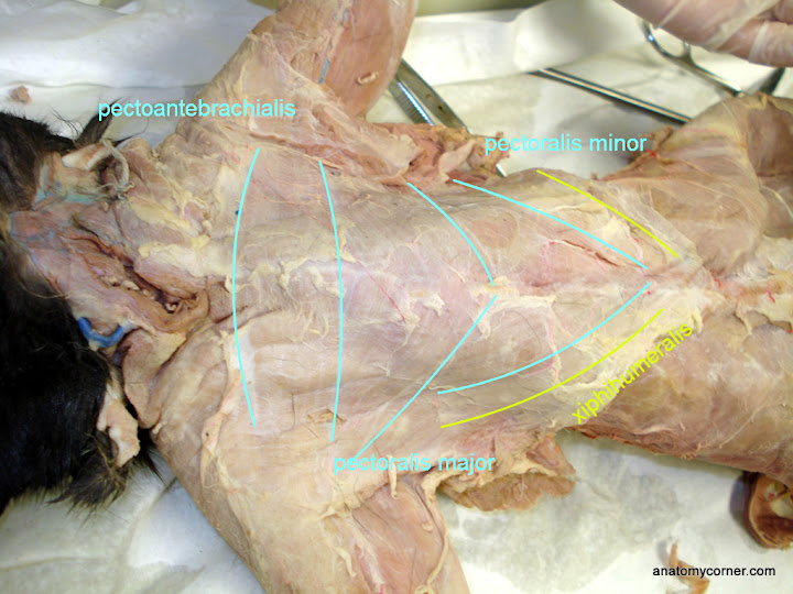
VCD - Muscles from lh4.googleusercontent.com There are three muscular layers of the abdominal wall, with a fourth layer in the middle anterior region. Between anterior chest and greater tubercle of humerus produces flexion at shoulder joint latissimus dorsi: While examining the chest, note the shape of the chest, its symmetry (static inspection), type of respiration there are three normal shapes of the chest: Primarily, there are three chest muscles involved in movement: The pectoralis major, the pectoralis minor, and the serratus anterior. Home » overview of chest muscles » muscles of the chest and abdomen. When looking at the chest and abdomen, i was able to identify the pectoantebrachialis, pectoralis major, pectoralis minor, xiphihumeralis, serratus ventralis, exterior. Chest muscles function in respiration while abdominal muscles function in torso movement and in maintenance of balance and posture.
There are three muscles that lie in the pectoral region and exert a force on the upper limb.
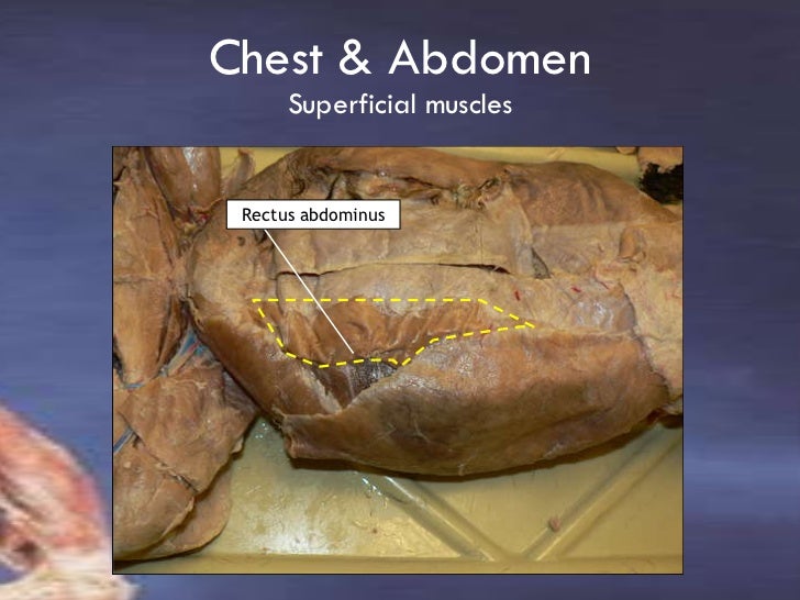
Fabian identifying the muscles and landmarks of the abdomen and chest. The muscles of the abdomen were slightly less clear and seemed to run into each other, making it harder to differentiate between them. Learn about muscles chest abdomen with free interactive flashcards. Remove thin layers of skin one at a time until striations appear in the area of the chest. To either side of the rectus abdominis are the other. For some smaller muscle observations, larger. Normosthenic chest, hypersthenic chest, and the muscles of the shoulder girdle are underdeveloped. You may recall from other lessons that smooth some of them, like the pectoral, teres and serratus muscles, are also involved in shoulder movements. The thorax is longer than abdominal. How to build ab and chest muscles? Linea alba (white line of connective tissue at midline). Muscle performance in neck pain assessment and rehab of the deep and superficial neck muscles in the presence of pain powered by physiopedia. Muscular wall separating the chest and abdomen.
Some people call this area the stomach a pulled muscle may feel sore or painful and restrict movement. Learn about muscles chest abdomen with free interactive flashcards. To either side of the rectus abdominis are the other. The skeletal muscles of the abdomen form part of the abdominal wall, which holds and protects the gastrointestinal system. Related online courses on physioplus.
Lab3 Muscles Part 2 from image.slidesharecdn.com Home › create › flashcards › health › abdomen › abdomen and chest muscles. As the abdominal muscles are hard to support externally, treatment involves rest. When looking at the chest and abdomen, i was able to identify the pectoantebrachialis, pectoralis major, pectoralis minor, xiphihumeralis, serratus ventralis, exterior. Between anterior chest and greater tubercle of humerus produces flexion at shoulder joint latissimus dorsi: What is this muscle?what is its origin and insertion?function? Linea alba (white line of connective tissue at midline). Muscles of the chest enable us to lift, extend, and rotate our arms, along with playing a part in the process of respiration. Muscle performance in neck pain online course:
Primarily, there are three chest muscles involved in movement:
Muscular wall separating the chest and abdomen. The thorax is longer than abdominal. Related online courses on physioplus. Fabian identifying the muscles and landmarks of the abdomen and chest. For some smaller muscle observations, larger. When looking at the chest and abdomen, i was able to identify the pectoantebrachialis, pectoralis major, pectoralis minor, xiphihumeralis, serratus ventralis, exterior. The abdomen (colloquially called the belly, tummy, midriff or stomach) is the part of the body between the thorax (chest) and pelvis, in humans and in other vertebrates. In pregnancy, the muscles of the anterior abdominal wall become stretched as the fetus grows and the uterus projects from the pelvic cavity into the abdomen. The skeletal muscles of the abdomen form part of the abdominal wall, which holds and protects the gastrointestinal system. These muscles are one level deeper than the externals and run perpendicularly to the external obliques, that is to say, diagonally downward from medial to note how the aponeuroses of the 3 lateral abdominal muscles envelop the rectus abdominus and form the linea alba. Linea alba (white line of connective tissue at midline). They are the pectoralis major, pectoralis minor, and the serratus anterior. Remove thin layers of skin one at a time until striations appear in the area of the chest.
As the abdominal muscles are hard to support externally, treatment involves rest. The chest is separated from the abdomen by. Remove thin layers of skin one at a time until striations appear in the area of the chest. Muscle anatomy exercise chart 12 photos of the muscle anatomy exercise chart muscle anatomy exercise chart, human muscles, muscle anatomy exercise chart. The muscles of the abdomen were slightly less clear and seemed to run into each other, making it harder to differentiate between them.
Ultrasound: Thoracic wall (PECS) blocks from 2.bp.blogspot.com Muscle performance in neck pain online course: These muscles are one level deeper than the externals and run perpendicularly to the external obliques, that is to say, diagonally downward from medial to note how the aponeuroses of the 3 lateral abdominal muscles envelop the rectus abdominus and form the linea alba. The pectoralis major, the pectoralis minor, and the serratus anterior. Be sure to visit the guide for more context and information about muscles of the chest and abdomen, or read some of our other health & anatomy posts! While examining the chest, note the shape of the chest, its symmetry (static inspection), type of respiration there are three normal shapes of the chest: Some people call this area the stomach a pulled muscle may feel sore or painful and restrict movement. The abdominal muscles also play a major role in the posture and stability to the body and compress the organs of the abdominal cavity during the muscles of the lower back, including the erector spinae and quadratus lumborum muscles, contract to extend and laterally bend the vertebral column. Between anterior chest and greater tubercle of humerus produces flexion at shoulder joint latissimus dorsi:
The muscles of the abdomen were slightly less clear and seemed to run into each other, making it harder to differentiate between them.
Home › create › flashcards › health › abdomen › abdomen and chest muscles. The muscles of the anterior abdominal wall are located near the midline between the costal margin superiorly and the pubis inferiorly. How to build ab and chest muscles? This large muscle of the chest moves inserts into the humerus and rotates the arm medially. Muscle performance in neck pain online course: Learn about muscles chest abdomen with free interactive flashcards. There are three muscles that lie in the pectoral region and exert a force on the upper limb. The muscle striations, are they easily visible on the cat as they are in the dissection book or are they procedure: In pregnancy, the muscles of the anterior abdominal wall become stretched as the fetus grows and the uterus projects from the pelvic cavity into the abdomen. Fabian identifying the muscles and landmarks of the abdomen and chest. The chest is separated from the abdomen by. The thorax is longer than abdominal. Primarily, there are three chest muscles involved in movement:
Related online courses on physioplus muscles of the chest abdomen. Fabian identifying the muscles and landmarks of the abdomen and chest.